

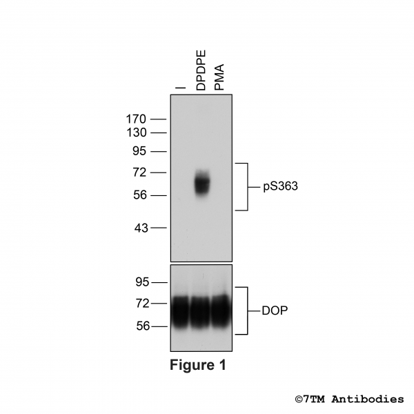
pS363-DOP (phospho-∂-Opioid Receptor Antibody)

Citations


- Order number: 7TM0317B
- Content: 100 µl
- Host: Rabbit


Citations

NEW

NEW

Citations

Citations

Citations

Citations
KO-Validated
, µ-Opioid Receptor Antibody")
Citations

Citations

Citations

Recently viewed