

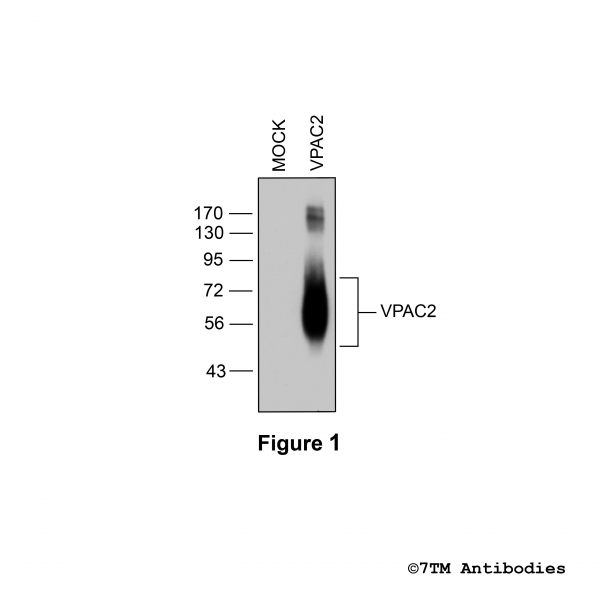
VPAC2 (non-phospho), VIP Receptor 2 Antibody

Citations
KO-Validated


- Order number: 7TM0372N
- Content: 100 µl
- Host: Rabbit


NEW

NEW

NEW
")
Citations
KO-Validated

NEW

NEW

NEW

Citations

Citations

Recently viewed