The CRF1 receptor antibody is directed against the distal part of the carboxyl-terminal tail of human CRF1. It can be used to detect total CRF1 receptors in Western blots independent of phosphorylation. The CRF1 antibody can also be used to isolate and enrich CRF1 receptors from cell and tissue lysates. It also detects CRF1 in cultured cells and tissue sections by immunohistochemistry.
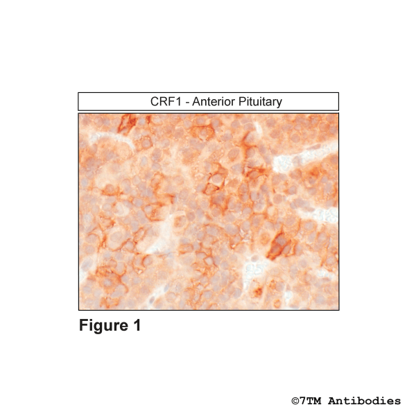
Figure 1. Immunohistochemical identification of Corticotropin-Releasing Factor Receptor 1 in anterior pituitary. Sections were dewaxed, microwaved in citric acid, and incubated with anti-CRF1 (Corticotropin-Releasing Factor Receptor 1) antibody (7TM0212N-IC) at a dilution of 1:100. Sections were then sequentially treated with biotinylated anti-rabbit IgG and avidin-biotin solution.Color was developed by incubation in 3-amino-9-ethylcarbazole (AEC), and sections were counterstained with hematoxylin. Note, CRF1 receptors were detected at the plasma membrane of a subpopulation of anterior pituitary cells.
Figure 2. Validation of the Corticotropin-Releasing Factor Receptor 1 in transfected HEK293 cells. Native HEK293 cells (MOCK) or HEK293 cells stably expressing the Corticotropin-Releasing Factor Receptor 1 (CRF1) were lysed and immunoblotted with the phosphorylation-independent anti-CRF1 antibody (7TM0212N-IC) at a dilution of 1:1000.
Figure 3. Immunocytochemical identification of Corticotropin-Releasing Factor Receptor 1 in HEK293 cells. HEK293 cells stably expressing the Corticotropin-Releasing Factor Receptor 1 (CRF1) were either not exposed or exposed to 100 nM CRF for 30 min and immunocytochemically stained with anti-CRF1 antibody (7TM0212N-IC) at a dilution of 1:200. Note, CRF1 receptors were confined to the plasma membrane in untreated cells (0 min).CRF1 receptors were seen in perinuclear clusters of vesicles after 30 min CRF exposure.