The FFA2 receptor antibody is directed against the distal part of the carboxyl-terminal tail of human FFA2. It can be used to detect total FFA2 receptors in Western blots independent of phosphorylation. The FFA2 antibody can also be used to isolate and enrich FFA2 receptors from cell and tissue lysates. It also detects FFA2 in cultured cells and tissue sections by immunohistochemistry.
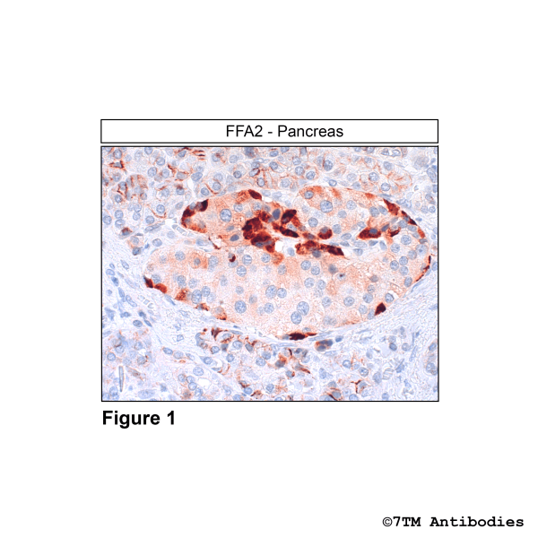
Figure 1. Immunohistochemical identification of FFA2 Receptor in pancreas. Sections were dewaxed, microwaved in citric acid, and incubated with anti-FFA2 (FFA2 Receptor) antibody (7TM0226N-IC) at a dilution of 1:100. Sections were then sequentially treated with biotinylated anti-rabbit IgG and avidin-biotin solution.Color was developed by incubation in 3-amino-9-ethylcarbazole (AEC), and sections were counterstained with hematoxylin. Note, FFA2 receptors were detected in a subset of pacreatic islet cells and at th eplasma membrane of exocrine pancreas cells.
Figure 2. Immunohistochemical identification of FFA2 Receptor in liver. Sections were dewaxed, microwaved in citric acid, and incubated with anti-FFA2 (FFA2 Receptor) antibody (7TM0226N-IC) at a dilution of 1:100. Sections were then sequentially treated with biotinylated anti-rabbit IgG and avidin-biotin solution.Color was developed by incubation in 3-amino-9-ethylcarbazole (AEC), and sections were counterstained with hematoxylin. Note, FFA2 receptors were detected at the plasma membrane of hepatocytes.
Figure 3. Immunohistochemical identification of FFA2 Receptor in colon carcinoma. Sections were dewaxed, microwaved in citric acid, and incubated with anti-FFA2 (FFA2 Receptor) antibody (7TM0226N-IC) at a dilution of 1:100. Sections were then sequentially treated with biotinylated anti-rabbit IgG and avidin-biotin solution.Color was developed by incubation in 3-amino-9-ethylcarbazole (AEC), and sections were counterstained with hematoxylin. Note, FFA2 receptors were detected at the plasma membrane of colon carcinoma cells.
Figure 4. Validation of the FFA2 in transfected HEK293 cells. Flp-In TREx HEK293 cells were induced to express human FFA2 DREADD-eYFP were treated with vehicle or 100 nM sorbate for 5 min. FFA2 receptors were enriched using a GFP tra and immunoblotted with the phosphorylation-independent anti-FFA2 antibody (7TM0226N-IC) at a dilution of 1:1000.