Prices plus VAT plus shipping costs
Ready to ship today,
Delivery time appr. 5-8 days
- Order number: 7TM0NPS-IC
- Content: 100 µl
- Host: Rabbit
The NPS antibody is directed against human Neuropeptide S. The NPS antibody can be used to detect Neuropeptide S in formalin-fixed, paraffin-embedded tissue sections by immunohistochemistry.
| Alternative Names | NPS, neuropeptide S |
| IUPHAR Target ID | 1490 |
| UniProt ID | P0C0P6 |
| Immunohistochemistry (IHC) | 1:100 |
| Western Blot (WB) | - |
| Species Reactivity | Human |
| Host / Isotype | Rabbit / IgG |
| Class | Polyclonal |
| Immunogen | A synthetic peptide presents human NPS. |
| Form | Liquid |
| Purification | Antigen affinity chromatography |
| Storage buffer | Dulbecco's PBS, pH 7.4, with 150 mM NaCl, 0.02% sodium azide |
| Storage conditions | short-term 4°C, long-term -20°C |
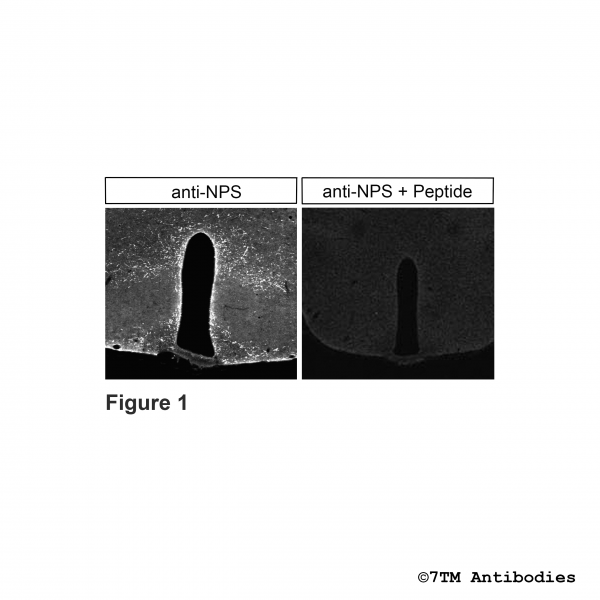
Figure 1. Immunohistochemical control of anti-NPS (Neuropeptide S) antibody in mouse coronal sections. Sections were quenched, blocked and incubated with anti-NPS (Neuropeptide S) antibody (7TM0NPS-IC) at a dilution of 1:100 (left picture) or incubated with the same antibody in the same dilution but antibody was preincubated with 1 µM Neuropeptide S (right picture). Sections were then washed and treated with Alexa Fluor 555-conjugated goat anti-rabbit IgG. Note, NPS staining was abolished in section when antibody was preincubated with NPS peptide.
Figure 2. Immunohistochemical identification of Neuropeptide S (NPS) in mouse telencephalon. Sections were quenched, blocked and incubated with anti-NPS (Neuropeptide S) antibody (7TM0NPS-IC) at a dilution of 1:100. Sections were then washed and treated with Alexa Fluor 555-conjugated goat anti-rabbit IgG. Note, strong NPS signals were detected in the medial part of the medial preoptic nucleus and moderate staining was observed in strial part and anterodorsal part of preoptic area.
Figure 3. Immunohistochemical identification of Neuropeptide S (NPS) in mouse diencephalon. Sections were quenched, blocked and incubated with anti-NPS (Neuropeptide S) antibody (7TM0NPS-IC) at a dilution of 1:100. Sections were then washed and treated with Alexa Fluor 555-conjugated goat anti-rabbit IgG. Note, NPS was detected in anterior part of the paraventricular thalamic nucleus. Right picture show NPS staining around A14 dopamine cells and anterodorsal thalimic nucleus.


