

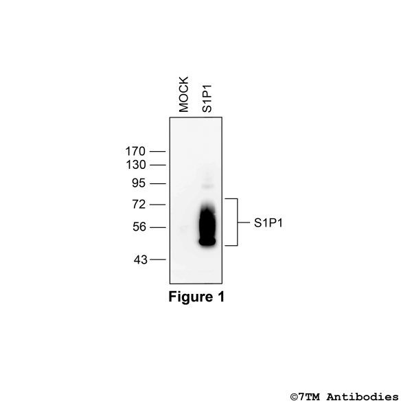
S1P1 (non-phospho), Sphingosine 1-Phosphate Receptor 1 Antibody

NEW


- Order number: 7TM0275N
- Content: 100 µl
- Host: Rabbit


NEW

NEW

NEW

NEW

NEW

NEW

NEW

NEW

NEW

NEW

NEW

NEW

Recently viewed