


Prices plus VAT plus shipping costs
Ready to ship today,
Delivery time appr. 5-8 days
- Order number: 7TM0075N
- Content: 100 µl
- Host: Rabbit


The non-phospho-XCR1 receptor antibody is directed against the distal part of the carboxyl-terminal tail of human XCR1. It can be used to detect total XCR1 receptors in Western blots independent of phosphorylation. The non-phospho-XCR1 antibody can also be used to isolate and enrich XCR1 receptors from cell and tissue lysates. It also detects XCR1 in cultured cells and tissue sections by immunohistochemistry.
| Alternative Names | CCXCR1, chemokine XC receptor 1, C motif-1/lymphotactin receptor |
| IUPHAR Target ID | 75 |
| UniProt ID | P46094 |
| Western Blot (WB) | 1:1000 |
| Immunohistochemistry (IHC) | 1:100 |
| Species Reactivity | Human |
| Host / Isotype | Rabbit / IgG |
| Class | Polyclonal |
| Immunogen | A synthetic peptide presents the carboxyl-terminal tail of human XCR1. |
| Form | Liquid |
| Purification | Antigen affinity chromatography |
| Storage buffer | Dulbecco's PBS, pH 7.4, with 150 mM NaCl, 0.02% sodium azide |
| Storage conditions | short-term 4°C, long-term -20°C |
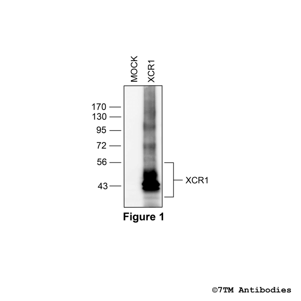
Figure 1. Validation of the XCR Chemokine Receptor 1 in transfected HEK293 cells. Native HEK293 cells (MOCK) or HEK293 cells stably expressing the XCR Chemokine Receptor 1 (XCR1) were lysed and immunoblotted with the phosphorylation-independent anti-XCR1 antibody (7TM0075N) at a dilution of 1:1000.
Figure 2. Immunohistochemical identification of XCR Chemokine Receptor 1 in pancreas. Sections were dewaxed, microwaved in citric acid, and incubated with anti-XCR1 (non-phospho-XCR Chemokine Receptor 1) antibody (7TM0075N) at a dilution of 1:100. Sections were then sequentially treated with biotinylated anti-rabbit IgG and avidin-biotin solution. Color was developed by incubation in 3-amino-9-ethylcarbazole (AEC), and sections were counterstained with hematoxylin.
Figure 3. Immunohistochemical varification of XCR Chemokine Receptor 1 antibody in pancreas. Sections were dewaxed, microwaved in citric acid, and not exposed (left pannel) or exposed to peptide (right pannel) that was used for production of anti-XCR1 (non-phospho-