


 in rat hypothalamus")
 in rat hypothalamus")

Prices plus VAT plus shipping costs
Ready to ship today,
Delivery time appr. 5-8 days
- Order number: 7TM0NPB-IC
- Content: 100 µl
- Host: Rabbit


The NPB antibody is directed against human Neuropeptide B. The NPB antibody can be used to detect Neuropeptide B in formalin-fixed, paraffin-embedded tissue sections by immunohistochemistry.
| Alternative Names | NPB, neuropeptide B |
| IUPHAR Target ID | 1502 |
| UniProt ID | Q8NG41 |
| Immunohistochemistry (IHC) | 1:100 |
| Western Blot (WB) | - |
| Species Reactivity | Human, Mouse, Rat |
| Host / Isotype | Rabbit / IgG |
| Class | Polyclonal |
| Immunogen | A synthetic peptide presents human NPB. |
| Form | Liquid |
| Purification | Antigen affinity chromatography |
| Storage buffer | Dulbecco's PBS, pH 7.4, with 150 mM NaCl, 0.02% sodium azide |
| Storage conditions | short-term 4°C, long-term -20°C |
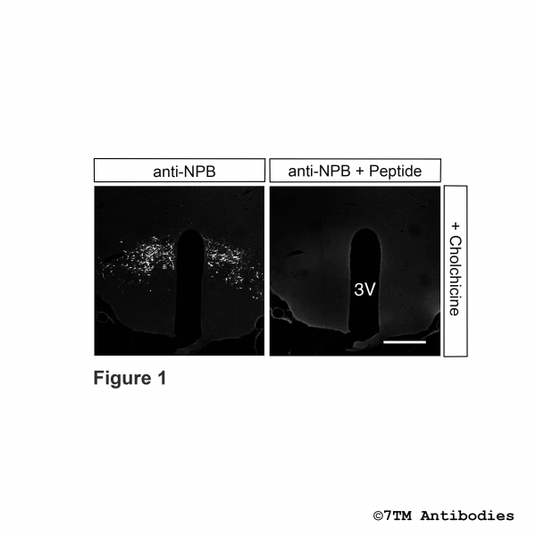
Figure 1. Immunohistochemical identification of Neuropeptide B (NPB) in rat hypothalamus. Rats were treated with an intracerebroventricular injection with 100 µg colchicine (10µg/µl) followed by vascular perfusion and fixation. Sections were dewaxed, microwaved in citric acid, and incubated with anti-NPB (Neuropeptide B) antibody (7TMNPB-IC) at a dilution of 1:100 or incubated with the same antibody in the same dilution but antibody was preincubated with 10 µg/ml Neuropeptide B (right picture). Sections were then sequentially treated with biotinylated anti-rabbit IgG and avidin-biotin solution. Color was developed by incubation with streptavidine-cyanine 3.18. Note, after colchicine treatement Neuropeptide B was located in neuronal somata. NPB staining was abolished in section when antibody was preincubated with NPB peptide.
Figure 2. Immunohistochemical identification of Neuropeptide B (NPB) in rat hypothalamus. Rats were treated with an intracerebroventricular injection of a vehicle instead of colchicine (Figure 1) followed by vascular perfusion and fixation. Sections were dewaxed, microwaved in citric acid, and incubated with anti-NPB (Neuropeptide B) antibody (7TMNPB-IC) at a dilution of 1:100 or incubated with the same antibody in the same dilution but antibody was preincubated with 10 µg/ml Neuropeptide B (right picture). Sections were then sequentially treated with biotinylated anti-rabbit IgG and avidin-biotin solution. Color was developed by incubation with streptavidine-cyanine 3.18. Note, without colchicine treatment Neuropeptide B was located in fibers and terminals. NPB staining was abolished in section when antibody was preincubated with NPB peptide.
.
Schulz S, Stumm R, Höllt V. Immunofluorescent identification of neuropeptide B-containing nerve fibers and terminals in the rat hypothalamus. Neurosci Lett. 2007 Jan 3;411(1):67-71. doi: 10.1016/j.neulet.2006.10.015. Epub 2006 Oct 25. PMID: 17067739.
 antibody in mouse coronal sections")
