Prices plus VAT plus shipping costs
Ready to ship today,
Delivery time appr. 5-8 days
- Order number: 7TM0014-SP
- Content: 7 x 20 µL
- Host: Rabbit
M2 Sample Pack consisting of all six available phospho- and one non-phospho-M2 Receptor Antibodies 7 x 20 µL trial size each. Specifically, this sample pack contains the following antibodies pT271-M2 (7TM0014A), pS282/pS283-M2 (7TM0014B), pS286/pT287/pS288-M2 (7TM0014C), pT307/pS309-M2 (7TM0014D), pT310/pS311-M2 (7TM0014E), pS315/pS320-M2 (7TM0014F) and M2 (non-phospho) (7TM0014N).
| Alternative Names | AChR M2, Chrm 2, Cholinergic Receptor muscarinic 2 |
| IUPHAR Target ID | 14 |
| UniProt ID | P08172 |
| Western Blot (WB) | 1:1000 |
| Immunocytochemistry (ICC) | - |
| Species Reactivity | Human |
| Host / Isotype | Rabbit / IgG |
| Class | Polyclonal |
| Immunogen | Synthetic phospho- and non-phospho peptides derived from human M2. |
| Form | Liquid |
| Purification | Antigen affinity chromatography |
| Storage buffer | Dulbecco's PBS, pH 7.4, with 150 mM NaCl, 0.02% sodium azide |
| Storage conditions | short-term 4°C, long-term -20°C |
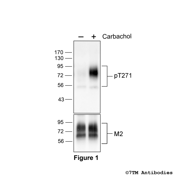
Figure 1. Agonist-induced Threonine271 phosphorylation of the M2 Muscarinic Acetycholine Receptor. Upper panel, HEK293 cells stably expressing the M2 Muscarinic Acetycholine Receptor (M2) were either not exposed or exposed to 100 nM Carbachol for 30 minutes. Cells were lysed and immunoblotted with the anti-pT271-M2 antibody (7TM0014A) at a dilution of 1:1000. Lower panel, blot was stripped and reprobed with an phosphorylation-independent anti-M2 antibody to confirm equal loading of the gel.
Figure 2. Agonist-induced Serine282/Serine283 phosphorylation of the M2 Muscarinic Acetylcholine Receptor. Upper panel, HEK293 cells stably expressing the M2 Muscarinic Acetylcholine Receptor (M2) were either not exposed or exposed to 100 nM Carbachol for 30 minutes. Cells were lysed and immunoblotted with the anti-pS282/pS283-M2 antibody (7TM0014B) at a dilution of 1:1000. Lower panel, blot was stripped and reprobed with an phosphorylation-independent anti-M2 antibody to confirm equal loading of the gel.
Figure 3. Agonist-induced Serine286/Threonine287/Serine288 phosphorylation of the M2 Muscarinic Acetycholine Receptor. Upper panel, HEK293 cells stably expressing the 5-Hydroxytryptamine Receptor 4 (M2) were either not exposed or exposed to 100 µM carbachol for 30 minutes. Cells were lysed and immunoblotted with the anti-pS286/pT287/pS288-M2 antibody (7TM0014C) at a dilution of 1:1000. Lower panel, blot was stripped and reprobed with an phosphorylation-independent anti-M2 antibody to confirm equal loading of the gel.
Figure 4. Agonist-induced Threonine307/Serine309 phosphorylation of the M2 Muscarinic Acetylcholine Receptor. Upper panel, HEK293 cells stably expressing the M2 Muscarinic Acetylcholine Receptor (M2) were either not exposed or exposed to 100 nM Carbachol for 30 minutes. Cells were lysed and immunoblotted with the anti-pT307/pS309-M2 antibody (7TM0014D) at a dilution of 1:1000. Lower panel, blot was stripped and reprobed with an phosphorylation-independent anti-M2 antibody to confirm equal loading of the gel.
Figure 5. Agonist-induced Threonine310/Serine311 phosphorylation of the M2 Muscarinic Acetycholine Receptor. Upper panel, HEK293 cells stably expressing the M2 Muscarinic Acetylcholine Receptor (M2) were either not exposed or exposed to 100 nM Carbachol for 30 minutes. Cells were lysed and immunoblotted with the anti-pT310/pS311-5-HT4 antibody (7TM0014E) at a dilution of 1:1000. Lower panel, blot was stripped and reprobed with an phosphorylation-independent anti-M2 antibody to confirm equal loading of the gel.
Figure 6. Agonist-induced Serine315/Serine320 phosphorylation of the M2 Muscarinic Acetycholine Receptor. Upper panel, HEK293 cells stably expressing the M2 Muscarinic Acetycholine Receptor (M2) were either not exposed or exposed to 100 nM Carbachol for 30 minutes. Cells were lysed and immunoblotted with the anti-pS315/pS320-5-HT4 antibody (7TM0014F) at a dilution of 1:1000. Lower panel, blot was stripped and reprobed with an phosphorylation-independent anti-M2 antibody to confirm equal loading of the gel.
Figure 7. Validation of the M2 Muscarinic Acetylcholine Receptor in transfected HEK293 cells. Native HEK293 cells (MOCK) or HEK293 cells stably expressing the M2 Muscarinic Acetylcholine Receptor (M2) were lysed and immunoblotted with the phosphorylation-independent anti-M2 antibody (7TM0014N) at a dilution of 1:1000.
Be the first to decipher the M2 phosphorylation barcode and let us know.


