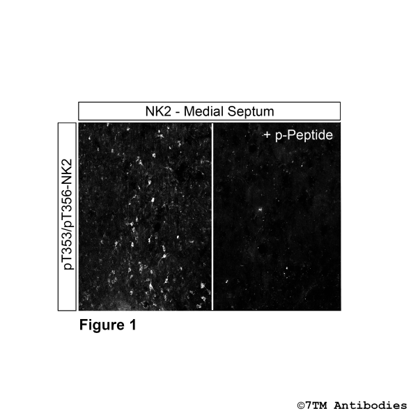
Prices plus VAT plus shipping costs
Ready to ship today,
Delivery time appr. 5-8 days
- Order number: 7TM0361C-IC
- Content: 300 µl
- Host: Rabbit
Threonine353/Threonine356 (T353/T356) is a major phosphorylation site of the NK2 receptor. The pT353/pT356-NK2 antibody detects phosphorylation in response to high-efficacy agonists. T353/T356 phosphorylation is a key regulator of NK2 desensitization, β-arrestin recruitment and internalization. pT353/pT356-NK2 antibody can detect phosphorylated NK2 in mouse brain in vivo.
Alternative Names | NK2, SKR, TACR2, Neurokinin B Receptor, Substance K Receptor |
IUPHAR Target ID | 361 |
UniProt ID | P21452 |
Immunohistochemistry (IHC) | 1:50 |
Species Reactivity | Human |
Host / Isotype | Rabbit / IgG |
Class | Polyclonal |
Immunogen | A synthetic phosphopeptide derived from human NK2 around the phosphorylation site of Thr353/Thr356. |
Form | Liquid |
Purification | Antigen affinity chromatography |
Storage buffer | Dulbecco's PBS, pH 7.4, with 150 mM NaCl, 0.02% sodium azide |
Storage conditions | short-term 4°C, long-term -20°C |
Figure 1. Immunohistochemical identification of Threonine353/Threonine356 phosphorylation of NK2 Receptor in Medial Septum. Sections were dewaxed, microwaved in citric acid, and not exposed (left pannel) or exposed to phospho-peptide (right pannel) that was used for production of anti-pT353/pT3565-NK2 (NK2 Receptor) antibody (7TM0361C-IC). Sections were then incubated with pT353//pT356-NK2 (NK2 Receptor) antibody (7TM0361C-IC) at a dilution of 1:100 and sequentially treated with biotinylated anti-rabbit IgG and avidin-biotin solution. Sections were then developed in 3,3-diaminobenzidine (DAB)-glucose oxidase and lightly counterstained with hematoxylin. Note, only in sections without peptide incubation NK2 receptors were uniformly detected at the plasma membrane of nearly all cells.
Figure 2. Agonist-induced Threonine353/Threonine356 phosphorylation of NK2 Receptor. Upper panel, HEK293 cells stably expressing the NK2-Receptor (NK2) were either not exposed or exposed to 1 µM of specific NK2-Receptor agonist Neurokinin B for 30 minutes. Cells were lysed and immunoblotted with the anti-pT353/pT356-NK2 antibody (7TM0361C-IC) at a dilution of 1:1000. Lower panel, blot was stripped and reprobed with an phosphorylation-independent anti-NK2 antibody to confirm equal loading of the gel.
Fritzwanker S, Nagel F, Kliewer A, Stammer V, Schulz S. In situ visualization of opioid and cannabinoid drug effects using phosphosite-specific GPCR antibodies. Commun Biol. 2023 Apr 15;6(1):419. doi: 10.1038/s42003-023-04786-2. PMID: 37061609; PMCID: PMC10105690.